Recognizing Symptoms of Rare Eye Diseases
Understanding the signs and symptoms of rare eye diseases is crucial for early diagnosis and effective management. Early recognition can help preserve vision and maintain a better quality of life.
Common Symptoms of Rare Eye Diseases
Rare eye diseases can present a variety of symptoms. Being aware of these can prompt timely medical consultation.
Gradual Vision Loss
- Description: A slow decline in vision that may not be immediately noticeable.
- Impact: Difficulty in performing daily activities like reading or driving.
Difficulty Seeing in Low Light
- Description: Trouble seeing in dimly lit environments or during nighttime.
- Impact: Increased risk of accidents and reduced ability to navigate in the dark.
Loss of Peripheral (Side) Vision
- Description: Narrowing of the field of vision, leading to tunnel vision.
- Impact: Challenges in detecting objects or movement outside the central vision.
Blurred or Distorted Vision
- Description: Objects may appear fuzzy, wavy, or misshapen.
- Impact: Difficulty in recognizing faces, reading, or performing detailed tasks.
Eye Pain or Discomfort
- Description: Persistent pain, aching, or a feeling of pressure in the eyes.
- Impact: Can interfere with concentration and overall comfort.
Increased Sensitivity to Light (Photophobia)
- Description: Unusually high sensitivity to bright lights or glare.
- Impact: Discomfort in well-lit environments and need to squint or wear sunglasses indoors.
Specific Symptoms of Key Rare Eye Diseases
Different rare eye diseases may have unique symptoms alongside the common ones.
Retinitis Pigmentosa
- Primary Symptoms:
- Night blindness
- Progressive loss of peripheral vision
- Reduced central vision over time
- Additional Signs: Difficulty adapting to changes in lighting.
Leber's Congenital Amaurosis
- Primary Symptoms:
- Severe vision loss or blindness at birth or in early infancy
- Nystagmus (involuntary eye movements)
- Enlarged pupils that do not respond well to light
- Additional Signs: Poor visual tracking and light sensitivity.
Other Rare Eye Conditions
- Choroideremia: Progressive loss of vision due to degeneration of the choroid.
- Stargardt Disease: Juvenile macular degeneration causing central vision loss.
- Usher Syndrome: Combination of vision and hearing loss.
When to Consult a Healthcare Professional
If you or a loved one experience any of the following, it’s important to seek medical advice:
- Sudden changes in vision
- Persistent eye pain
- Difficulty seeing in low light
- Gradual loss of peripheral vision
- Blurry or distorted vision
- Unusual light sensitivity
Early consultation can lead to timely diagnosis and management, potentially slowing disease progression.
Diagnosis and Treatment Options
Diagnosing rare eye diseases typically involves several specialized tests:
- Comprehensive Eye Exams: To assess overall eye health and vision clarity.
- Electroretinography (ERG): Measures the electrical responses of the retina.
- Genetic Testing: Identifies specific genetic mutations associated with the disease.
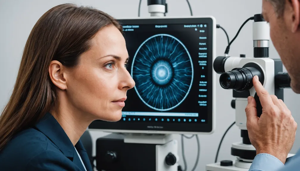
- Optical Coherence Tomography (OCT): Provides detailed images of the eye’s structures.
Treatment Options: - Medications: To manage symptoms or slow disease progression. - Vision Aids: Such as specialized glasses or low-vision devices. - Gene Therapy: Emerging treatments targeting specific genetic mutations. - Support Services: Including counseling and support groups to assist with adaptation.
Healthcare Professionals Who Can Assist
Managing rare eye diseases often requires a multidisciplinary approach. The following specialists can provide comprehensive care:
- Ophthalmologists: Medical doctors specializing in eye and vision care, capable of performing eye surgery and diagnosing eye diseases.
- Optometrists: Eye care professionals who perform eye exams and prescribe corrective lenses.
- Genetic Counselors: Assist in understanding genetic aspects of eye diseases and family planning.
- Retinal Specialists: Ophthalmologists with expertise in diseases of the retina.
- Low Vision Therapists: Help patients make the most of their remaining vision through training and adaptive techniques.
- Vision Rehabilitation Therapists: Support individuals in adapting to vision loss and maintaining independence.
Living with a Rare Eye Disease
Coping with a rare eye disease involves both medical treatment and emotional support. Here are some strategies to help manage daily life:
- Stay Informed: Understanding your condition helps in making informed decisions about your care.
- Seek Support: Connect with support groups or counselors to share experiences and gain emotional support.
- Use Assistive Technology: Tools like screen readers, magnifiers, and specialized software can enhance independence.
- Maintain a Healthy Lifestyle: Regular exercise, a balanced diet, and avoiding smoking can support overall health.
- Plan for the Future: Consider legal and financial planning to ensure security as the disease progresses.
Recognizing the symptoms of rare eye diseases and seeking timely medical attention can make a significant difference in managing these conditions effectively.
