Diagnostic Imaging: MRI, CT Scans, and PET Scans Explained
When facing a rare cancer of the nervous system, understanding the tools doctors use to diagnose and monitor your condition is essential. Diagnostic imaging plays a crucial role in identifying the location, size, and nature of tumors in the brain, spinal cord, or peripheral nerves. This lesson will help you grasp the basics of MRI, CT scans, and PET scans, and how they contribute to your care.
Understanding Diagnostic Imaging
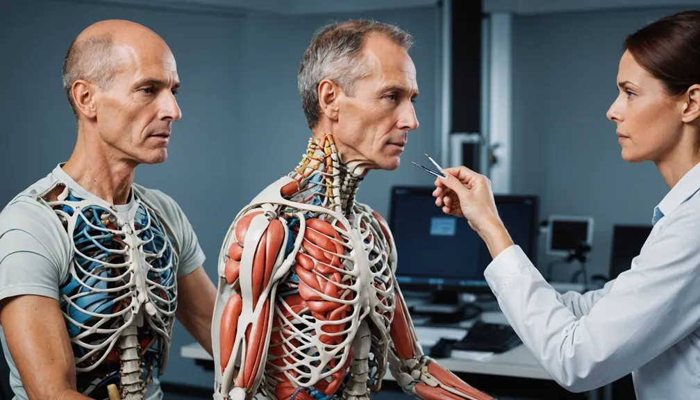
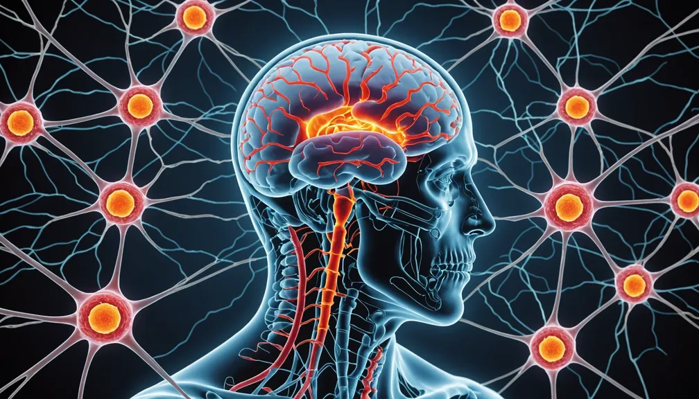
Diagnostic imaging refers to a variety of techniques used to create visual representations of the interior of your body. These images help doctors detect abnormalities, guide treatments, and monitor the progress of diseases like rare nervous system cancers.
MRI (Magnetic Resonance Imaging)
What is an MRI?
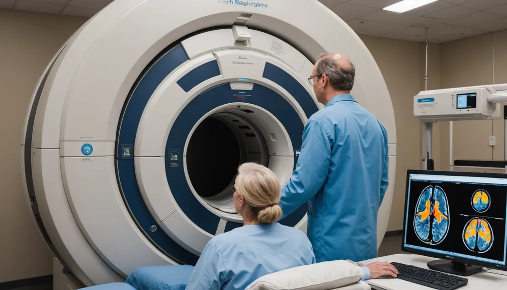
An MRI uses strong magnetic fields and radio waves to produce detailed images of organs and tissues within the body, particularly useful for examining the nervous system.
How does an MRI work?
- Magnetic Fields: The MRI machine generates powerful magnets that align the protons in your body.
- Radio Waves: Pulses of radio waves disturb this alignment.
- Image Creation: As the protons return to their original position, they emit signals that are converted into detailed images by a computer.
What to expect during an MRI
- Preparation: You may be asked to remove metal objects and change into a hospital gown.
- Procedure: You'll lie down on a sliding table that moves into the MRI machine. The process is painless but can last between 30 to 60 minutes.
- Comfort Tips: Relax and remain still to ensure clear images. You might wear earplugs to reduce noise from the machine.
Benefits of MRI for Diagnosing Nervous System Cancers
- High Detail: Provides clear images of soft tissues, making it easier to identify tumors.
- No Radiation: Unlike CT scans, MRIs do not use ionizing radiation.
- Versatility: Can assess the brain, spinal cord, and peripheral nerves effectively.
CT Scan (Computed Tomography Scan)
What is a CT Scan?
A CT scan combines multiple X-ray images taken from different angles to create cross-sectional views of the body, offering more detailed information than standard X-rays.
How does a CT Scan work?
- X-Rays: The CT machine emits X-rays that pass through your body.
- Image Processing: Detectors measure the X-rays, and a computer assembles them into detailed images.
- Contrast Agents: Sometimes, a contrast dye is used to highlight specific areas, enhancing image clarity.
What to expect during a CT Scan
- Preparation: You may need to drink a contrast liquid or receive it through an IV.
- Procedure: You'll lie on a table that moves through the scanner while X-rays are taken. The scan typically takes about 10 to 30 minutes.
- Comfort Tips: Remain still to ensure accurate images. Inform your doctor if you feel uncomfortable or claustrophobic.
Benefits of CT Scans for Diagnosing Nervous System Cancers
- Speed: CT scans are quick, making them suitable for urgent situations.
- Bone Detail: Excellent for visualizing bone structures and detecting fractures or bone involvement by tumors.
- Versatility: Useful for guiding biopsies and planning surgeries.
PET Scan (Positron Emission Tomography Scan)
What is a PET Scan?
A PET scan is a type of nuclear medicine imaging that shows how your tissues and organs are functioning, often used to detect cancerous cells.
How does a PET Scan work?
- Radioactive Tracers: A small amount of radioactive sugar is injected into your bloodstream.
- Cell Activity: Cancer cells, which consume more sugar, absorb more of the tracer.
- Image Creation: The PET scanner detects the tracer’s radiation and creates images showing areas of high metabolic activity.
What to expect during a PET Scan
- Preparation: Fasting for several hours before the scan may be required.
- Procedure: You'll receive an injection of the tracer and wait for it to circulate. Then, you'll lie down on a table that moves through the scanner. The entire process can take a few hours.
- Comfort Tips: Stay relaxed during the waiting period and inform the medical team if you feel uneasy.
Benefits of PET Scans for Diagnosing Nervous System Cancers
- Functional Imaging: Shows how tissues are working, which helps identify active cancer cells.
- Detection of Metastasis: Can reveal if cancer has spread to other parts of the body.
- Treatment Monitoring: Assesses how well treatments are working by measuring changes in metabolic activity.
Comparing MRI, CT, and PET Scans
Differences and Similarities
- MRI vs. CT: MRI provides better soft tissue contrast without radiation, while CT is faster and better for bone structures.
- PET vs. MRI/CT: PET offers functional information about cellular activity, complementing the structural details from MRI and CT.
- Combination: Often, these scans are used together to provide a comprehensive view of the cancer.
When Each Scan is Recommended
- MRI: Preferred for detailed images of the brain, spinal cord, and nerves.
- CT Scan: Chosen for quick imaging needs and bone evaluations.
- PET Scan: Utilized for detecting active cancer, checking for metastasis, and monitoring treatment effectiveness.
Preparing for Your Imaging Appointment
What You Need to Know
- Medical History: Inform your doctor about any medical conditions, allergies, or previous imaging studies.
- Medications: Discuss any medications you are taking, as some may require adjustments.
- Claustrophobia: Let your healthcare provider know if you have anxiety, so they can offer support or alternatives.
Tips to Make the Process Easier
- Comfortable Clothing: Wear loose-fitting clothes without metal fasteners.
- Stay Still: Practice staying calm and still during the scan for clear images.
- Ask Questions: Don’t hesitate to ask the technician about the procedure if you’re unsure.
After the Scan: Understanding Your Results
How to Interpret the Findings
- Report Review: Your doctor will explain the images and what they reveal about your cancer.
- Next Steps: Based on the results, your treatment plan may be adjusted or further tests may be recommended.
Next Steps in Your Care
- Treatment Planning: Imaging results help determine the best course of treatment, such as surgery, radiation, or chemotherapy.
- Follow-Up Scans: Regular imaging may be necessary to monitor your progress and detect any changes early.
Healthcare Professionals Who Can Help
Types of Doctors and Specialists
- Neurosurgeon: Specializes in surgical treatments for nervous system cancers.
- Oncologist: Focuses on cancer diagnosis and treatment plans.
- Radiologist: Expert in interpreting diagnostic imaging results.
Supportive Health Professionals
- Nurse Navigator: Assists with coordinating your care and appointments.
- Physical Therapist: Helps maintain or improve physical function and manage symptoms.
- Counselor or Psychologist: Provides emotional support and coping strategies.
Understanding the role of diagnostic imaging in your care can empower you to make informed decisions and actively participate in your treatment journey. Always feel free to discuss any questions or concerns with your healthcare team to ensure you receive the support and information you need.
