Diagnostic Tools: EKG, Echocardiogram, and MRI Explained
Understanding the diagnostic tools used to evaluate rare cardiovascular diseases can empower you and your loved ones to engage actively in your healthcare journey. This lesson covers three essential diagnostic methods: the Electrocardiogram (EKG), Echocardiogram, and Magnetic Resonance Imaging (MRI).
Electrocardiogram (EKG)
What is an EKG?
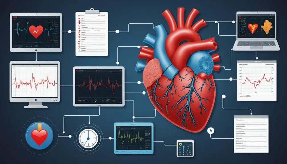
An Electrocardiogram (EKG) is a simple, painless test that records the electrical activity of your heart. It helps doctors assess how well your heart is functioning and detect any irregularities in its rhythm.
How is an EKG Performed?
- Preparation: No special preparation is needed. You may be asked to remove jewelry or clothing from the upper body.
- Procedure: Small, sticky patches called electrodes are placed on your chest, arms, and legs.
- Duration: The test typically takes about 5-10 minutes.
- Experience: You will lie still while the EKG machine records your heart's electrical activity. It is non-invasive and painless.
What Does an EKG Show?
- Heart Rate and Rhythm: Identifies irregular heartbeats, such as arrhythmias.
- Heart Size: Detects if the heart is enlarged.
- Electrical Conduction: Reveals issues with the heart's electrical pathways.
- Previous Heart Attacks: Can indicate if you've had a heart attack in the past.
Why is an EKG Important?
For individuals with rare cardiovascular diseases, an EKG is crucial for: - Early Detection: Identifying heart abnormalities before symptoms appear. - Monitoring Progress: Tracking changes in heart function over time. - Guiding Treatment: Assisting doctors in choosing the best treatment options.
Echocardiogram
What is an Echocardiogram?
An Echocardiogram is an ultrasound-based test that uses sound waves to create moving images of your heart. It provides detailed information about the structure and function of your heart.
Types of Echocardiograms
- Transthoracic Echocardiogram (TTE): The most common type, performed by moving a transducer over the chest.
- Transesophageal Echocardiogram (TEE): Involves inserting a specialized probe down the throat to get a closer look at the heart.
How is an Echocardiogram Performed?
- Preparation: Wear comfortable clothing and avoid wearing metal objects on your chest.
- Procedure: Gel is applied to your chest, and the transducer is moved to capture images.
- Duration: Typically lasts between 30 to 60 minutes.
- Experience: Non-invasive and painless, though TEE may require mild sedation.
What Does an Echocardiogram Show?
- Heart Chambers and Valves: Examines the size and movement of heart chambers and the function of valves.
- Heart Muscle Movement: Assesses how well the heart muscle is contracting and relaxing.
- Blood Flow: Evaluates the flow of blood through the heart and major vessels.
- Structural Abnormalities: Detects congenital heart defects or other structural issues.
Why is an Echocardiogram Important?
For those with rare cardiovascular conditions, an echocardiogram helps: - Diagnose Conditions: Identifying specific heart abnormalities. - Evaluate Severity: Understanding the extent of heart disease. - Monitor Treatment: Assessing how well treatments are working over time.
Magnetic Resonance Imaging (MRI)
What is a Cardiovascular MRI?
A Cardiovascular Magnetic Resonance Imaging (MRI) is a non-invasive imaging technology that uses powerful magnets and radio waves to create detailed images of the heart and blood vessels.
How is a Cardiovascular MRI Performed?
- Preparation: You may need to remove metal objects and inform your doctor if you have any implants.
- Procedure: You lie inside a large machine that makes loud noises during the scan.
- Duration: The scan can take anywhere from 30 minutes to over an hour.
- Experience: While the procedure is painless, it requires you to remain still. Some patients may feel claustrophobic.
What Does a Cardiovascular MRI Show?
- Heart Structure: Detailed images of the heart's anatomy, including chambers, walls, and valves.
- Blood Vessels: Visualizes blood flow and identifies blockages or abnormalities in arteries and veins.
- Tissue Health: Detects scarring, inflammation, or other tissue changes in the heart.
- Functional Assessment: Measures how well the heart pumps blood and how efficiently blood flows through the vessels.
Why is a Cardiovascular MRI Important?
For individuals with rare cardiovascular diseases, an MRI offers: - Comprehensive Evaluation: Provides a thorough assessment of the heart and blood vessels. - Accurate Diagnosis: Helps identify complex structural and functional issues. - Treatment Planning: Assists doctors in developing effective treatment strategies based on detailed imaging.
Healthcare Professionals Who Can Help
Managing rare cardiovascular diseases often requires a team of specialized healthcare providers. The following professionals may be involved in your care:
- Cardiologist: A doctor specializing in heart and blood vessel diseases, responsible for diagnosis and treatment.
- Cardiovascular Surgeon: A surgeon who performs operations on the heart and blood vessels.
- Radiologist: A medical doctor who interprets imaging tests like EKGs, echocardiograms, and MRIs.
- Genetic Counselor: Provides information and support if your condition has a genetic component.
- Nurse Specialist: Offers care coordination, education, and support throughout your treatment.
- Physical Therapist: Helps you recover and maintain physical function, especially after procedures.
- Primary Care Physician: Manages overall health and coordinates care among specialists.
Engaging with these professionals ensures a comprehensive approach to managing your rare cardiovascular disease, utilizing the appropriate diagnostic tools to guide effective treatment.


