Diagnostic Procedures: Pulmonary Function Tests and Imaging
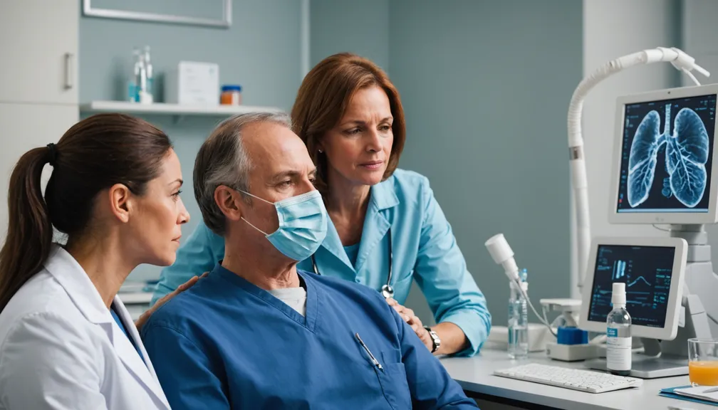
Understanding the diagnostic procedures for rare respiratory diseases can help you feel more prepared and empowered. This lesson covers Pulmonary Function Tests (PFTs) and Imaging Studies, explaining what they involve, how they assist in diagnosis, and what to expect during each procedure.
1. Pulmonary Function Tests (PFTs)
What Are Pulmonary Function Tests?
Pulmonary Function Tests are a group of tests that measure how well your lungs are working. They assess lung volume, capacity, rates of flow, and gas exchange.
Types of Pulmonary Function Tests
- Spirometry: Measures how much air you can inhale and exhale, and how quickly you can exhale.
- Gas Diffusion Tests: Assess how well oxygen passes from your lungs into your blood.
- Plethysmography: Measures the total volume of air in your lungs after taking a deep breath.
- Exercise Testing: Evaluates lung function during physical activity.
Preparing for Pulmonary Function Tests
- Avoid Caffeine and Smoking: Do not consume caffeine or smoke for at least four hours before the test, as they can affect results.
- Medication Management: Inform your doctor about any medications you are taking, as some may need to be paused before the test.
- Wear Comfortable Clothing: Choose loose-fitting clothes to make breathing easier during the tests.
During the Test
- Spirometry Example:
- Breathing Normally: Start by breathing normally into the spirometer.
- Deep Breath: Take a deep breath and exhale as forcefully and quickly as possible.
-
Repeat: You may be asked to repeat the process several times to ensure accurate results.
-
Comfort and Safety: PFTs are non-invasive and generally safe, though you may feel slight discomfort during forceful breathing maneuvers.
Understanding Your PFT Results
- Normal vs. Abnormal Results: Your doctor will compare your results to standard values based on age, gender, height, and ethnicity.
- Interpreting Data: Results can indicate restrictive or obstructive lung diseases, helping to narrow down possible conditions.
How PFTs Help in Diagnosing Respiratory Diseases
- Identifying Lung Function Issues: PFTs can detect early changes in lung function, even before symptoms appear.
- Guiding Treatment Plans: Results help healthcare providers choose the most effective treatments and monitor disease progression.
2. Imaging Studies
What Are Imaging Studies?
Imaging Studies create pictures of the structures inside your body, particularly the lungs and airways, to help diagnose respiratory conditions.
Common Imaging Techniques for Respiratory Diseases
- Chest X-Ray:
- Purpose: Provides a basic view of the lungs, heart, and chest bones.
-
Uses: Detects infections, fractures, and certain lung conditions.
-
High-Resolution Computed Tomography (HRCT):
- Purpose: Offers detailed images of the lung tissue.
-
Uses: Diagnoses interstitial lung diseases, such as idiopathic pulmonary fibrosis.
-
Magnetic Resonance Imaging (MRI):
- Purpose: Uses magnetic fields to produce detailed images.
-
Uses: Evaluates blood vessels and soft tissues in the chest.
-
Positron Emission Tomography (PET) Scan:
- Purpose: Shows how tissues and organs are functioning.
- Uses: Detects cancerous cells and assesses metabolic activity in the lungs.
Preparing for Imaging Procedures
- Fasting Requirements: Some tests may require you to avoid eating or drinking for a few hours beforehand.
- Clothing and Accessories: Wear comfortable, metal-free clothing, and remove jewelry to prevent interference with imaging equipment.
- Inform About Allergies: Let your healthcare provider know if you have any allergies, especially to contrast materials used in some scans.
Understanding Your Imaging Results
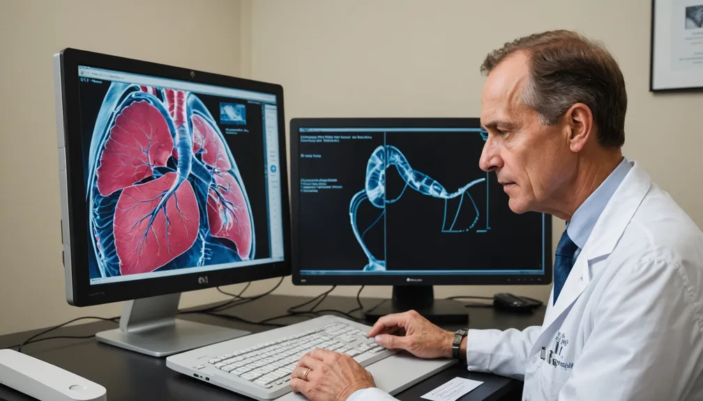
- Report Interpretation: A radiologist will analyze the images and provide a report to your doctor, highlighting any abnormalities.
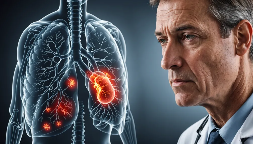
- Visual Insights: Images can reveal structural changes, inflammation, scarring, or tumors in the lungs and airways.
How Imaging Studies Aid in Diagnosis
- Locating Problems: Help identify the exact location and extent of lung damage or disease.
- Monitoring Progress: Allow doctors to track changes in your condition over time and adjust treatments accordingly.
3. The Diagnostic Team
Types of Doctors and Health Professionals Involved
- Pulmonologists: Specialists in lung and respiratory diseases who oversee diagnosis and treatment.
- Radiologists: Doctors trained to interpret imaging studies and provide detailed reports.
- Respiratory Therapists: Assist with breathing treatments and help perform pulmonary function tests.
- Nurses and Support Staff: Provide care, answer questions, and support you throughout the diagnostic process.
4. Tips for Patients and Caregivers
Preparing for Diagnostic Tests
- Gather Information: Write down your symptoms, medical history, and any questions you have before the appointment.
- Assist Each Other: Caregivers can help remember instructions and provide emotional support during tests.
Managing Anxiety During Tests
- Relaxation Techniques: Practice deep breathing, visualization, or listen to calming music to stay relaxed.
- Communicate with Your Team: Let your healthcare providers know if you’re feeling anxious—they can offer reassurance and adjustments as needed.
Communicating with Your Healthcare Team
- Ask Questions: Don’t hesitate to ask for clarification on procedures, results, or treatment options.
- Express Concerns: Share any worries or discomfort you experience during tests to receive the best possible care.
Remember: Early and accurate diagnosis through pulmonary function tests and imaging studies is essential in managing rare respiratory diseases. Working closely with your healthcare team can lead to better treatment outcomes and an improved quality of life.

